Most common x-ray techniques
X-ray images are one of the most common methods used to examine the mouth and teeth. Your dentist will decide whether x-rays are required, but the imaging itself can be done by a nurse or dental hygienist, provided they have had the training to do so. The most common x-ray techniques used in dentistry include periapical, bite-wing and panoramic x-rays. The types of x-ray imaging available may vary between our dental clinics. Where relevant, your dentist will discuss the type of x-rays needed with you at your appointment.
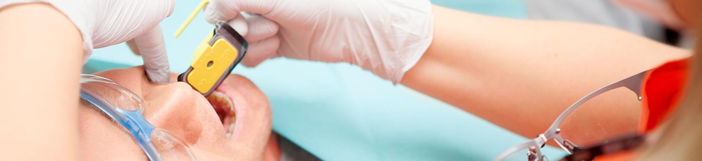
Periapical x-rays
Periapical x-rays are the most common type of x-ray imaging used by dentists. The x-ray film is placed in the patient’s mouth so that the site captured is located between the film and the x-ray tube. The radiation emanating from the x-ray will be absorbed by the object, generating an image. Periapical x-ray images include both the tooth and the root. It is used to diagnose caries or cavities, tooth infections and periodontitis.
Bite-Wing x-rays
A Bite-Wing x-ray allows dental professionals to investigate harder to reach areas inside the mouth, including the areas between your teeth and under any existing fillings. Often it can be used to identify caries, which causes cavities in your teeth. Unlike the periapical images, Bite-Wing x-rays do not show the teeth roots.
Other common x-ray techniques are panoramic x-rays and cone beam CT.
X-ray radiation exposure
The level of radiation used for dental x-ray purposes can vary significantly and depends on the area involved and the precision required. However, the amount of radiation you will be exposed to is low enough not to be associated with any adverse effects. That said, we will always do everything we can to avoid unnecessary exposure wherever possible and the dentist will always carry out a case-by-case assessment of whether x-rays are needed. However, if you have been exposed to radiation, due to recent medical investigations for example, please discuss this with your dentist beforehand.